38 using a microscope worksheet
Using a microscope. Subject: Biology. Age range: 11-14. Resource type: Worksheet/Activity. Rating: 4.6 · 31 reviews · Free · In stock In this lesson students identify parts of a microscope, use a microscope to ... of a microscope by completing the worksheet Colour the microscope parts.
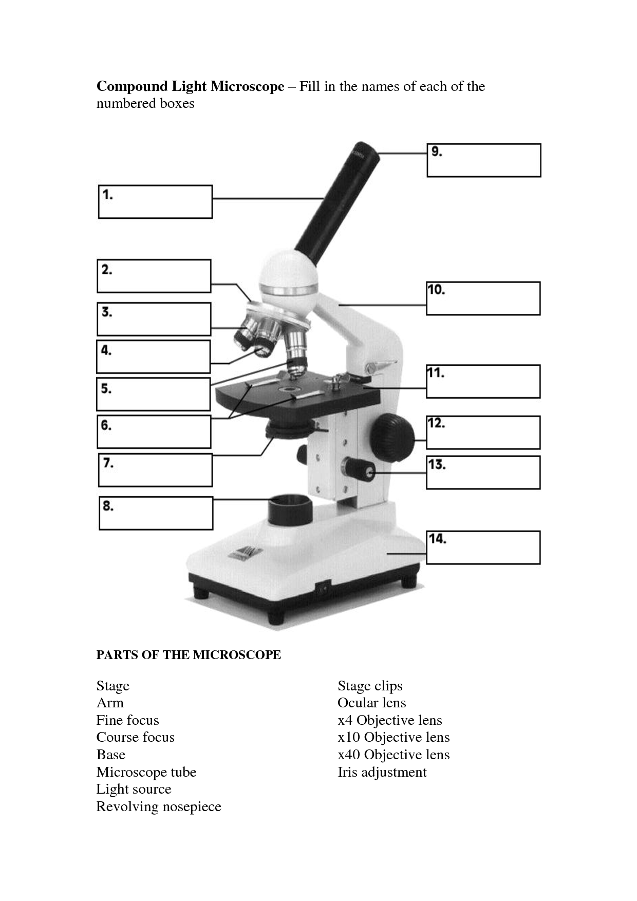
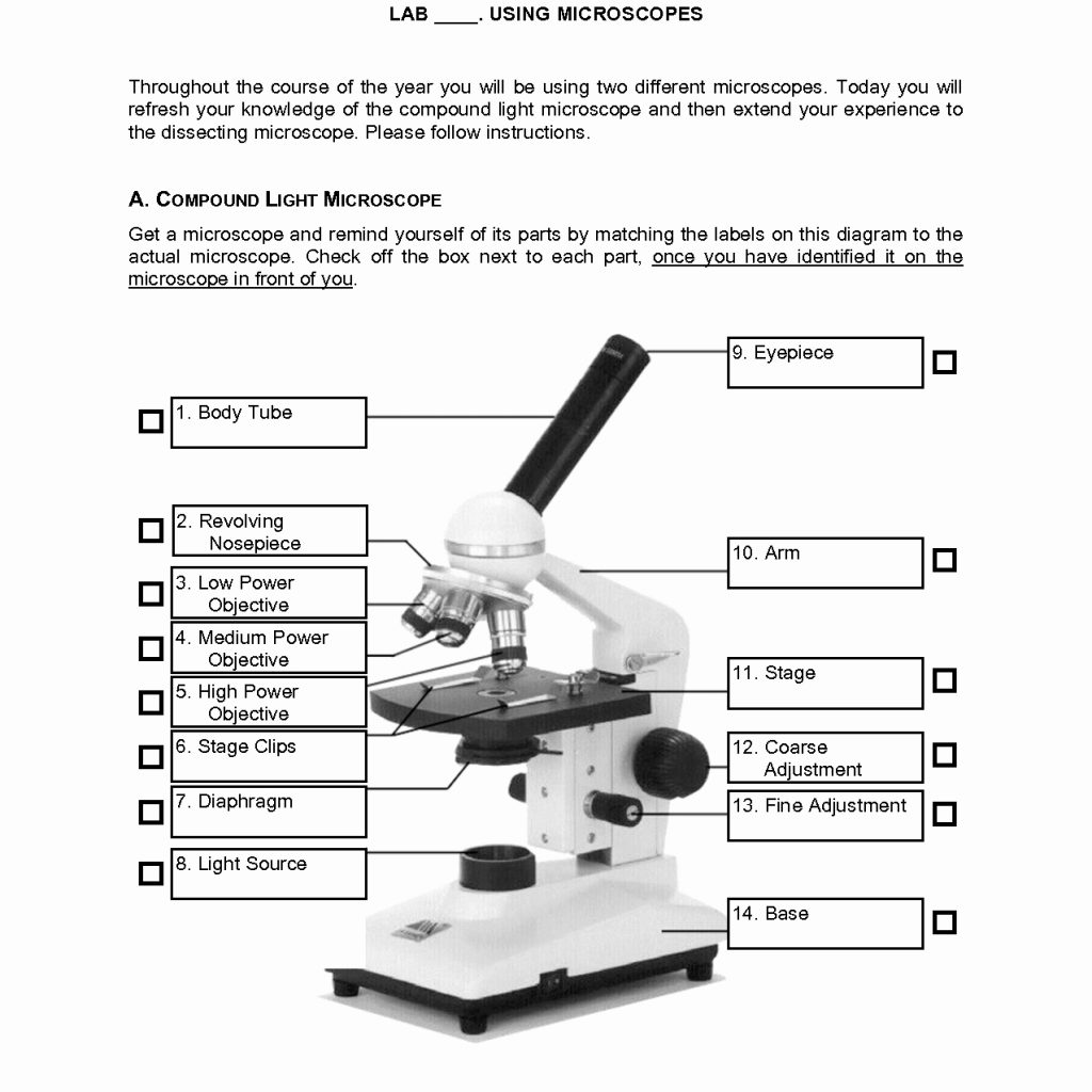
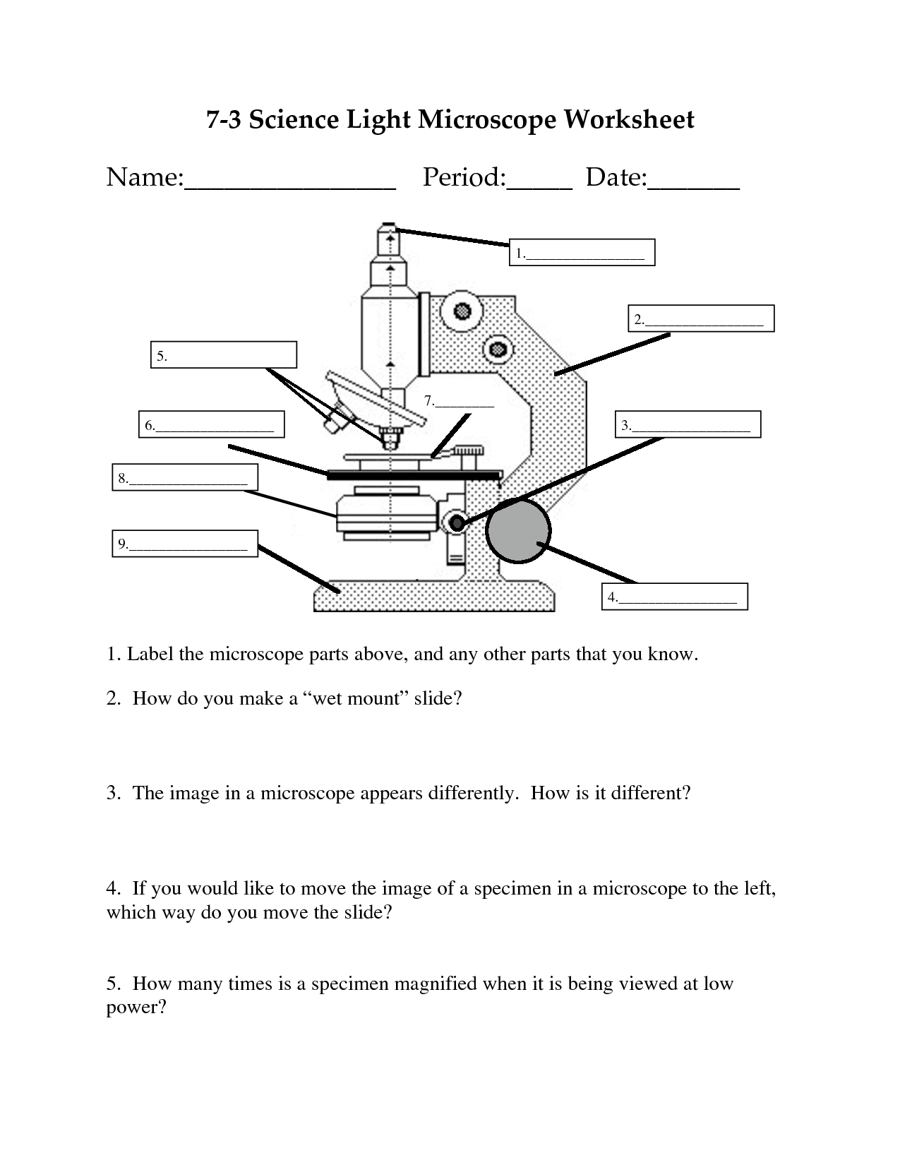
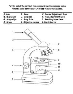
Before purchasing or using a microscope, it ... Eyepiece Lens: the lens at the top that you look through. ... Microscope Parts and Use Worksheet.3 pages

Using a microscope worksheet
In this worksheet, students will learn about the different parts of a light microscope, be able to label one correctly and learn how to use one. Doctors often use microscopes. Is there one in your doctor's office? Parts of the Microscope. Eyepiece: Lens closes to the eye.3 pages With the inclusion of a range of worksheets and teaching ideas, you will be completely prepared to begin the cells and organisations unit with our detailed ... Rating: 5 · 15 reviews
Using a microscope worksheet. Types of Microscopes. Light Microscope - the models found in most schools, use compound lenses to magnify objects. The lenses bend or refract light to make ... Results 1 - 24 of 1363 — Learning to use a microscope is an important part of science studies. With these Microscope Activity Worksheets, elementary students ... With the inclusion of a range of worksheets and teaching ideas, you will be completely prepared to begin the cells and organisations unit with our detailed ... Rating: 5 · 15 reviews Doctors often use microscopes. Is there one in your doctor's office? Parts of the Microscope. Eyepiece: Lens closes to the eye.3 pages
In this worksheet, students will learn about the different parts of a light microscope, be able to label one correctly and learn how to use one.
Microscopes Worksheet

How To Use A Microscope Worksheet - MOTHERSDAYGIFTSMOTHERSDAY

31 The Optical Microscope Worksheet 9 Answers - Free ...

Microscope Worksheet | Homeschooldressage.com
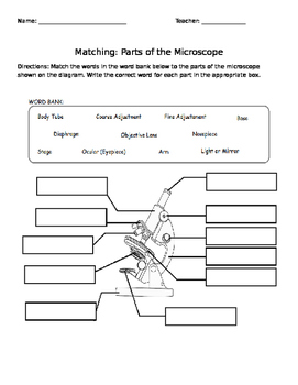
Microscope Labeling Worksheet | Free Worksheets Samples

The Compound Microscope Worksheet Answers - Micropedia

Microscope Worksheet Answers Quizlet - Worksheetpedia

Microscope Parts and Function Activities | Microscope ...

Parts Of A Microscope Worksheet | Homeschooldressage.com
Parts of the Microscope Worksheet by Amanda Behen | TpT

Microscope Worksheet Answer Key - Worksheetpedia

Printable Microscope Worksheet Pdf - Micropedia

Using A Microscope Worksheet

33 Microscope Parts And Use Worksheet Answer Key ...
This photo was made with some experimental liquids as milk, water paint and oil. I’ve made this with a friend and we had so much fun doing it. The surprise of the reactions thought the different material was both charming and changeling. I truly recommend everyone to try something like this, let’s share the different results. Have fun using this picture.
17 Best Images of Microscope Labeling Worksheet ...

Brainpop Microscope Worksheet Answers - Micropedia

Microscope Basicswkst
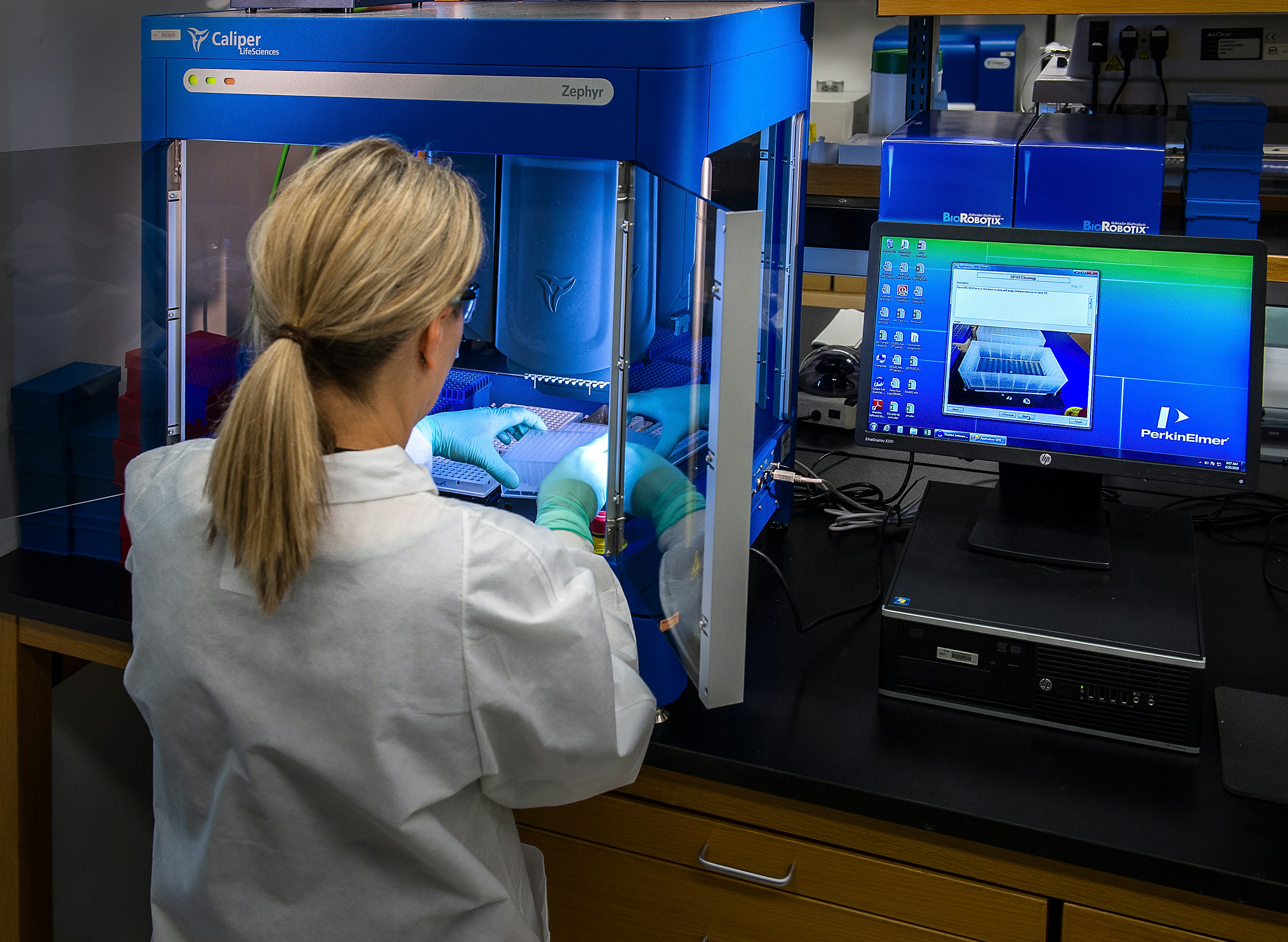
This image depicted a Centers for Disease Control and Prevention (CDC) scientist interacting with her Caliper LifeSciences’ Zephyr Molecular Biology Workstation, working with samples to be tested using a real-time PCR machine, known as a themocycler (see PHIL 22904), in order to identify the various types of poliovirus contained therein. The data from this analysis is stored in a computer, while the software further analyzes the data before being reviewed by a scientist.

Using A Microscope Worksheet

Using A Microscope Worksheet
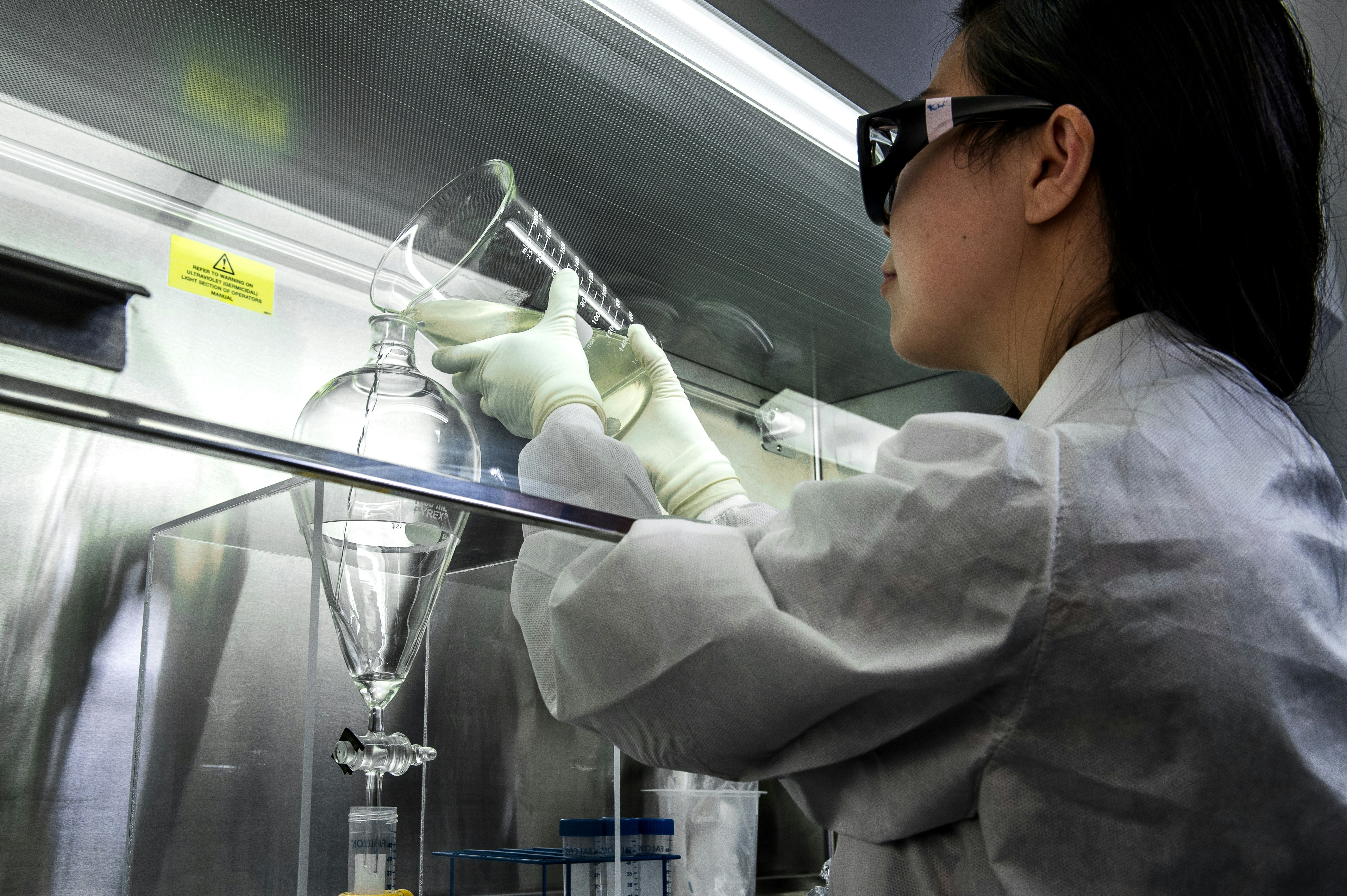
This photograph depicted a Centers for Disease Control and Prevention (CDC) scientist, concentrating poliovirus from sewage, so that the virus could be grown in cultured cells, and then tested using molecular methods. She was performing a polio environmental surveillance technique. There is no routine polio environmental surveillance in the U.S., but surveillance is done in many countries.
12 Best Images of Light Microscope Parts Worksheet ...
The Microscope Worksheets Answers

Using The Microscope Worksheet Answer Key - Best Bren
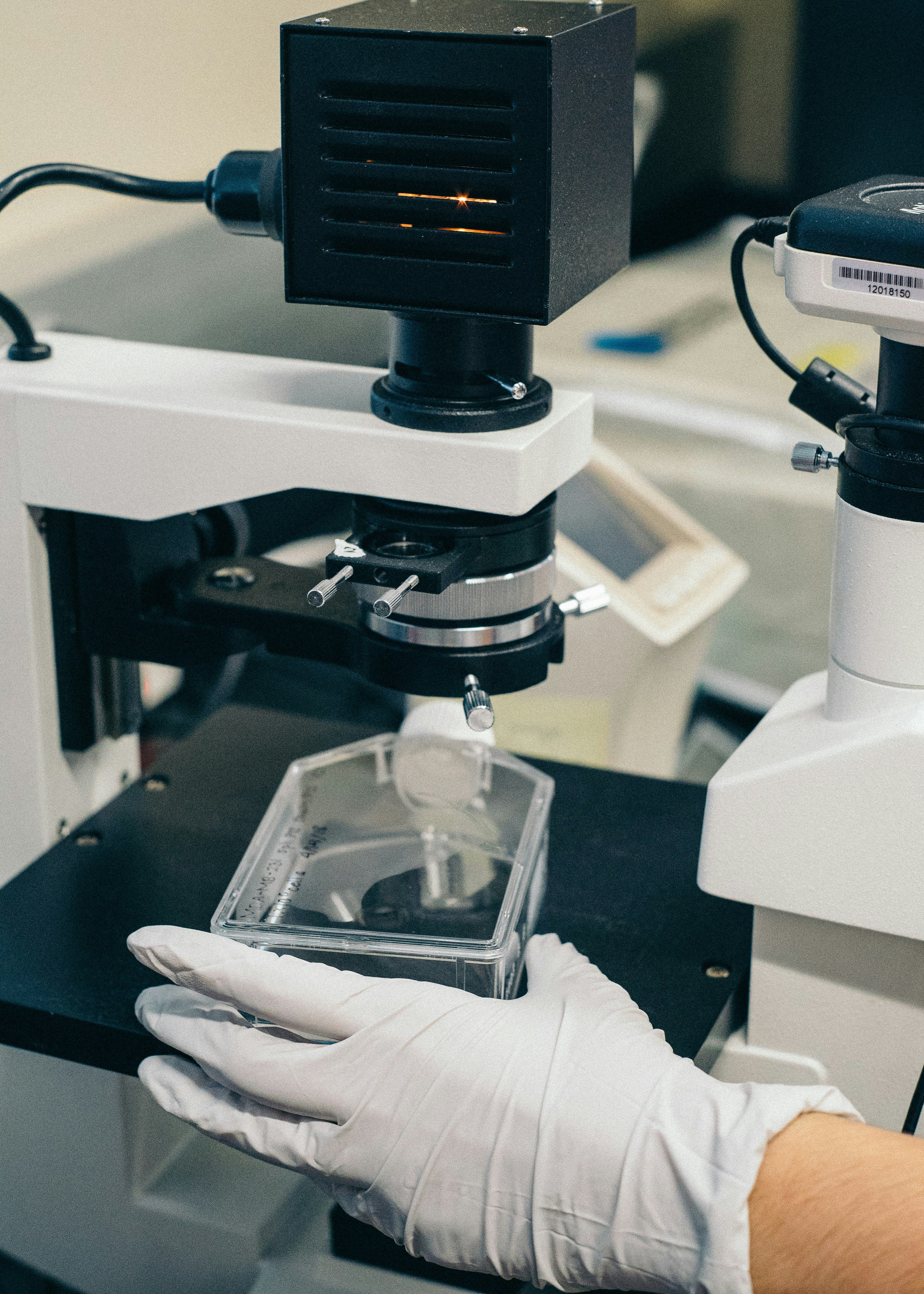
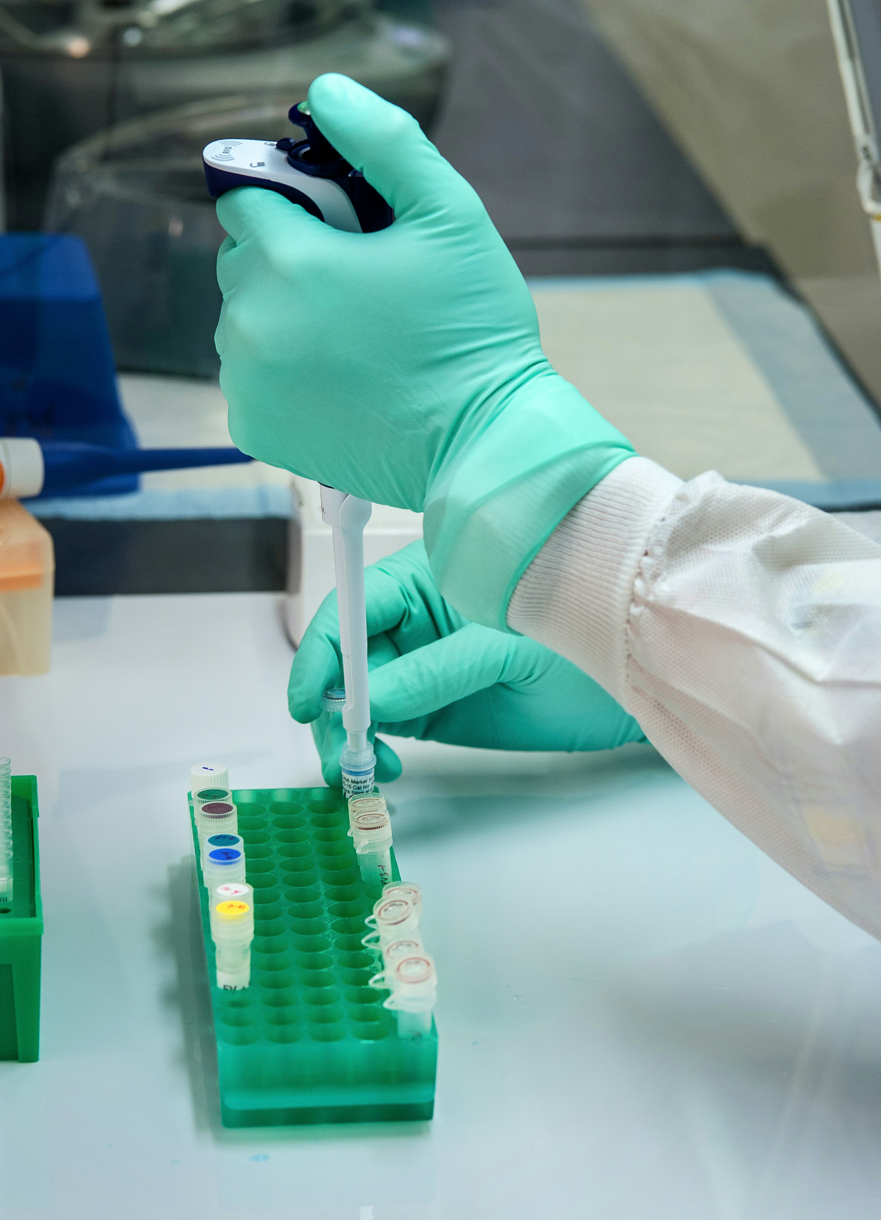
A gloved hand preparing a stem cell culture for analysis.
Parts of a Microscope Overview Reading Comprehension and ...

17 Best Images of Blank Microscope Worksheet - Blank ...
Microscope Worksheet For Middle School - Worksheet List

Cell Theory Worksheets & Definition - Laney Lee Teaching ...
The Compound Microscope Worksheet
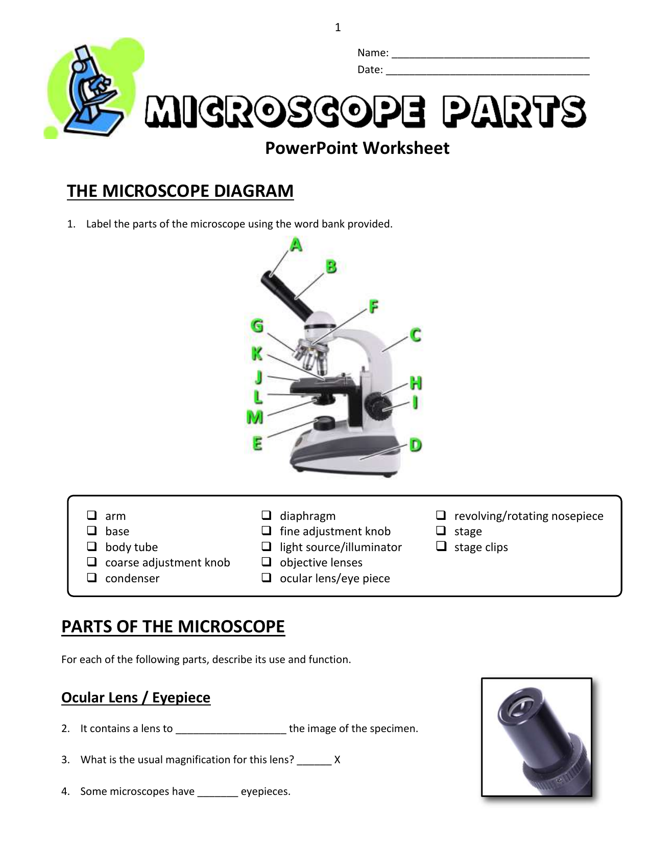
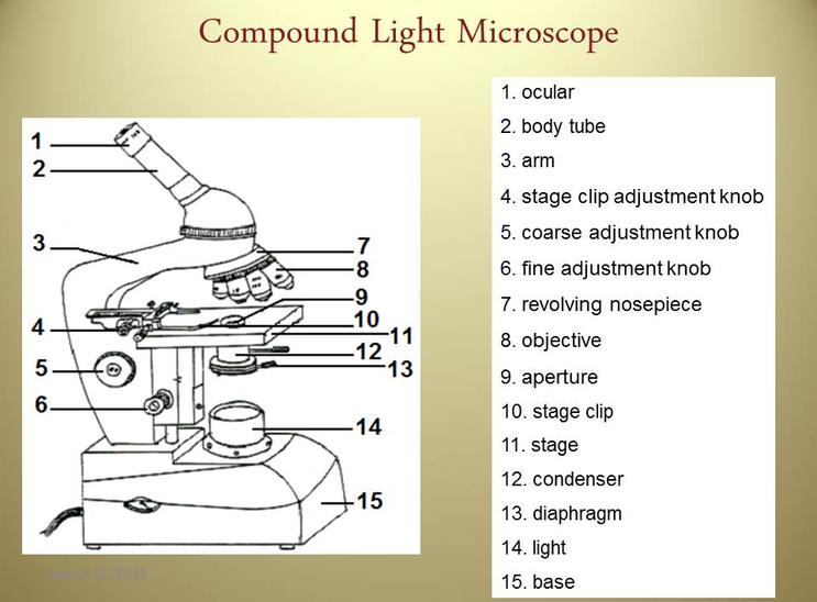
13 - Microscope Parts - PowerPoint Worksheet copy
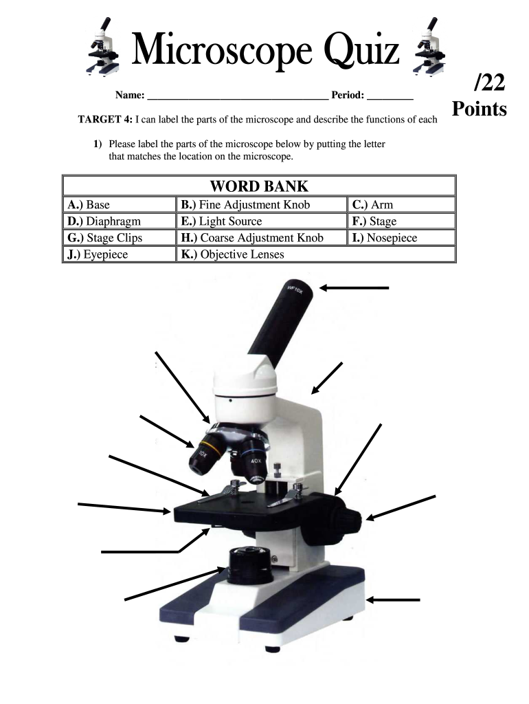
13 Microscope Parts Powerpoint Worksheet — db-excel.com
This Centers for Disease Control and Prevention (CDC) scientist was shown implementing molecular testing, in order to test for different types of polio. The 6-assay screening can determine which samples are polio, the specific serotype of polio, and whether they are vaccine, or wild strains.
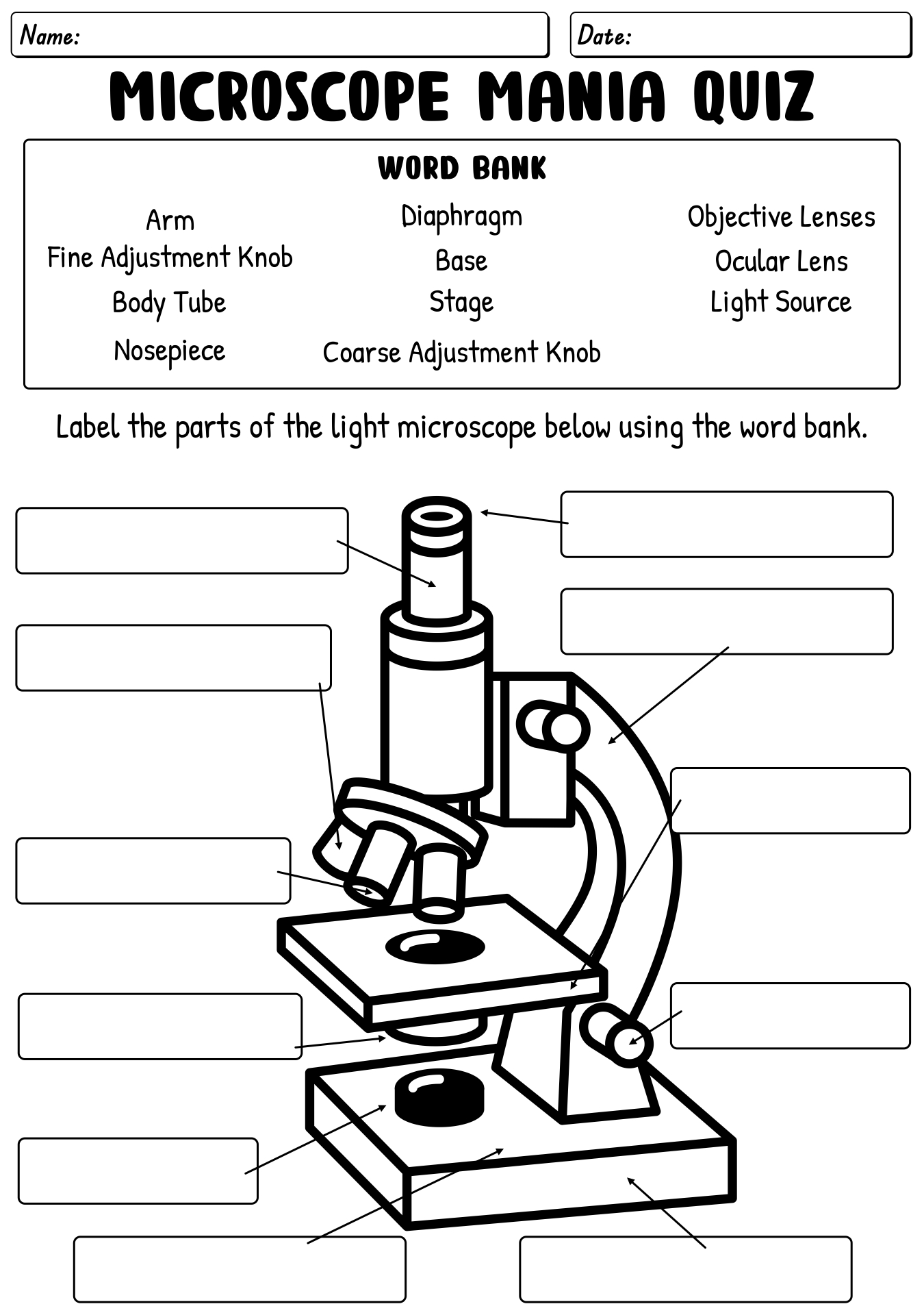
30 Microscope Mania Worksheet Answers - Worksheet Source 2021

Using Microscopes: Quiz & Worksheet for Kids | Study.com
Robert Charles Gallo, former Biomedical Researcher. He is best known for his work with the Human Immunodeficiency Virus (HIV), the infectious agent responsible for the Acquired Immune Deficiency Syndrome (AIDS). He was the former Chief of Laboratory of Tumor Cell Biology at the National Institutes of Health. 1980
Biomedical engineer uses microscope
0 Response to "38 using a microscope worksheet"
Post a Comment